How Blood Pumps Through Your Heart: Simple Flow Guide
Your heart works every second of every day, continuously moving blood through your lungs, organs, muscles, and tissues.
Although most people know that the heart “pumps blood,” the actual process is a carefully coordinated cycle involving four heart chambers, four valves, major blood vessels, the lungs, and an electrical conduction system.
Understanding how blood pumps through your heart can make it easier to understand blood pressure, oxygen circulation, heartbeats, heart valve problems, coronary artery disease, heart failure, and many other cardiovascular conditions.
Blood flows from your body to the right heart, moves to the lungs for oxygen, returns to the left heart, then pumps through the body again.
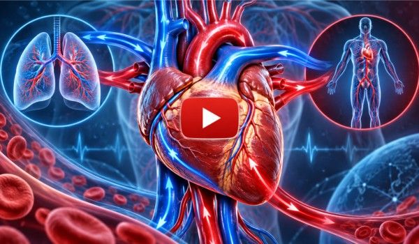

At its simplest, blood follows a continuous pathway: it returns from the body to the right side of the heart, travels to the lungs to receive oxygen, returns to the left side of the heart, and is then pumped back out to the rest of the body. But much more happens during every heartbeat.
This guide explains exactly how blood flows through the heart, what each chamber and valve does, why the right and left sides of the heart perform different jobs, and how the cardiovascular system keeps oxygen and nutrients circulating throughout your body.
What Does the Heart Actually Do?
The heart is a muscular organ located in the chest between the lungs. Its main responsibility is to keep blood circulating through the cardiovascular system.
Blood performs several essential jobs. It delivers oxygen from the lungs to tissues, transports nutrients, carries hormones and other chemical signals, and moves waste products to organs that can process or eliminate them.
For circulation to continue, blood must keep moving. The heart provides the force required to keep that movement going. Rather than functioning as a single hollow pump, the heart has four chambers that work together.
The chambers receive blood, move it through valves, pump it toward the lungs, and eventually send oxygen-rich blood throughout the entire body.
The heart also adjusts its pumping activity to meet your body’s needs. During sleep or quiet rest, the demand for oxygen is relatively low.
During exercise, physical work, stress, or other activities, the heart can beat faster and pump more blood to support increased oxygen requirements.
The Four Chambers of the Heart
To understand how blood pumps through your heart, you first need to understand its four chambers. The upper chambers are the atria, while the lower chambers are the ventricles.
🔷 Right Atrium
The right atrium is the first major chamber that receives blood returning from most of the body. This returning blood contains relatively less oxygen because tissues
Have already used much of the oxygen delivered during the previous circulation through the body. Large veins carry this blood into the right atrium, preparing it for the next stage of circulation.
🔷 Right Ventricle
Blood travels from the right atrium into the right ventricle. The right ventricle is responsible for pumping this oxygen-poor blood toward the lungs.
It does not need to generate of much pressure as the left ventricle because the lungs are relatively close to the heart. This part of circulation is called pulmonary circulation.
🔷 Left Atrium
After blood travels through the lungs and receives oxygen, it returns to the heart and enters the left atrium. The left atrium therefore receives oxygen-rich, not oxygen-poor, blood. From here, blood moves into the left ventricle.
🔷 Left Ventricle
The left ventricle is the heart’s primary pumping chamber for systemic circulation. It must generate enough force to push blood through the aorta and into arteries that eventually distribute blood throughout the entire body.
Because of this demanding workload, the muscular wall of the left ventricle is substantially thicker than that of the right ventricle.
How Blood Pumps Through Your Heart Step by Step
The easiest way to understand heart circulation is to follow one portion of blood through the complete cycle.
- Blood returns from the body. Oxygen-poor blood returns through large veins known as the superior vena cava and inferior vena cava and enters the right atrium.
- Blood enters the right ventricle. The right atrium moves blood through the tricuspid valve into the right ventricle.
- The right ventricle sends blood toward the lungs. When the right ventricle contracts, blood passes through the pulmonary valve and enters the pulmonary artery.
- Blood receives oxygen in the lungs. In the lungs, blood participates in gas exchange. Carbon dioxide moves from the blood to the air that will be exhaled, while oxygen enters the bloodstream.
- Oxygen-rich blood returns to the heart. The pulmonary veins carry the newly oxygenated blood from the lungs to the left atrium.
- Blood moves into the left ventricle. Blood passes through the mitral valve and fills the left ventricle.
- The left ventricle pumps blood into the aorta. When the left ventricle contracts, blood travels through the aortic valve into the aorta.
- Blood circulates throughout the body. The aorta branches into progressively smaller arteries that distribute oxygen-rich blood to organs, muscles, the brain, and other tissues. After delivering oxygen, the blood eventually returns through veins, and the entire cycle begins again.
This process occurs continuously throughout your life.
The Simple Blood Flow Pattern to Remember
If the anatomy seems complicated, remember this basic sequence:
Body → Right Atrium → Right Ventricle → Lungs → Left Atrium → Left Ventricle → Body
The right side of the heart primarily sends blood toward the lungs. The left side primarily sends blood throughout the body. The two sides work simultaneously, which makes circulation far more efficient than if blood had to move through the chambers one step at a time.
Why Does Blood Need to Travel to the Lungs?
Every cell in your body requires oxygen to support normal energy production and cellular function. As blood circulates through tissues, oxygen moves from the bloodstream into cells. The blood simultaneously collects carbon dioxide, a waste product associated with cellular metabolism.
That blood must then return to the lungs. Inside the lungs are microscopic air sacs called alveoli. These structures provide a large surface area for gas exchange between air and nearby blood vessels.
Carbon dioxide moves toward the lungs to be exhaled, while oxygen from inhaled air enters the blood. Once oxygenated, blood returns to the heart through the pulmonary veins.
The heart then sends it throughout the body again. This close relationship between the lungs and heart is one reason respiratory and cardiovascular health are so closely connected.
What Do the Four Heart Valves Do?
Moving blood is only part of the heart’s job. Blood also needs to travel in the correct direction. That is where heart valves become essential.
The heart contains four major valves: the tricuspid valve, pulmonary valve, mitral valve, and aortic valve. These structures work like carefully timed one-way doors.
When pressure changes inside the heart, the appropriate valve opens, allowing blood to move forward. As pressure changes again, the valve closes to help prevent significant backward flow.

🔷 Tricuspid Valve
The tricuspid valve is located between the right atrium and right ventricle. Blood passes through it while moving from the upper right chamber into the lower right chamber.
When the ventricle contracts, the valve closes, directing blood toward the pulmonary artery rather than backward into the atrium.
🔷 Pulmonary Valve
The pulmonary valve sits between the right ventricle and pulmonary artery. It opens when the right ventricle contracts and sends blood toward the lungs. The valve then closes as the ventricle relaxes.
🔷 Mitral Valve
The mitral valve is located between the left atrium and left ventricle. It allows oxygen-rich blood returning from the lungs to enter the powerful left ventricle. It also helps prevent blood from moving backward into the left atrium when the left ventricle contracts.
🔷 Aortic Valve
The aortic valve sits between the left ventricle and the aorta. It opens when the left ventricle contracts, allowing blood to enter the body’s largest artery.
It then closes, limiting blood from flowing backward into the ventricle. Problems affecting any of these valves can interfere with normal circulation.
Why Arteries Do Not Always Carry Oxygen-Rich Blood
One common misunderstanding is that arteries always carry oxygen-rich blood and veins always carry oxygen-poor blood. That is not quite correct.
Arteries are defined primarily by the direction blood travels: arteries carry blood away from the heart. Veins carry blood toward the heart.
Most arteries in systemic circulation carry oxygen-rich blood, but the pulmonary artery is a notable exception. It carries oxygen-poor blood from the right ventricle to the lungs.
Likewise, most veins carry blood that has relatively less oxygen, but pulmonary veins are exceptions. They bring oxygen-rich blood from the lungs back toward the left atrium. Remembering “arteries away, veins toward” can make cardiovascular anatomy much easier to understand.
What Makes Your Heart Pump?
Heart muscle does not contract randomly. The heart’s own electrical conduction system controls the timing of each heartbeat.
An electrical impulse normally begins in an area of specialized cells called the sinoatrial node, or SA node, located in the right atrium.
The SA node is often described as the heart’s natural pacemaker. The signal spreads through the atria, encouraging them to contract.
This helps move blood into the ventricles. The electrical signal then reaches another important structure called the atrioventricular node, or AV node.
After a short delay that helps the ventricles finish filling, electrical activity travels through specialized pathways into the ventricles. The ventricles then contract.
This synchronized electrical activity ensures that the atria and ventricles pump in the correct sequence rather than contracting chaotically. When the heart’s electrical signals become abnormal, an irregular heart rhythm, or arrhythmia, can develop.
What Happens During One Heartbeat?
Every heartbeat contains two broad phases: contraction and relaxation. The contraction phase is called systole. During ventricular systole, the ventricles contract.
The right ventricle pushes blood toward the lungs, while the left ventricle sends blood into the aorta and systemic circulation. The relaxation phase is known as diastole.
During this period, the ventricles relax and refill with blood in preparation for the next contraction. The process repeats again and again. This alternating pattern of filling and pumping creates the pulse that you can feel at places such as your wrist or neck.
What Creates the “Lub-Dub” Heart Sound?
The familiar heartbeat sound heard through a stethoscope is closely related to the closing of the heart valves.
The first sound, commonly described as “lub,” occurs primarily when the valves between the atria and ventricles close around the beginning of ventricular contraction.
The second sound, “dub,” is associated mainly with the closure of the aortic and pulmonary valves as the ventricles finish contracting.
Healthcare professionals can listen to heart sounds because unusual sounds may sometimes provide clues about abnormal blood flow or valve function.
For example, turbulent blood movement may create an additional sound called a heart murmur. Some murmurs are harmless, while others may indicate structural or valve abnormalities and require medical evaluation.
What Is Pulmonary Circulation?
Pulmonary circulation refers specifically to blood movement between the heart and lungs. It begins when the right ventricle pumps oxygen-poor blood into the pulmonary artery.
The pulmonary arteries divide into progressively smaller blood vessels as they approach the lungs. Eventually, blood reaches tiny capillaries located near the alveoli.
Gas exchange occurs here. The newly oxygenated blood then travels through pulmonary veins and returns to the left atrium. Pulmonary circulation operates at a lower pressure than systemic circulation because blood only needs to travel between the heart and nearby lungs.
What Is Systemic Circulation?
Systemic circulation begins when the left ventricle sends blood into the aorta. From there, arteries branch repeatedly and distribute blood toward different regions of the body.
As arteries become smaller, they eventually lead into microscopic blood vessels known as capillaries. Capillaries are where much of the exchange between blood and tissues takes place.
Oxygen and nutrients can move toward cells, while substances such as carbon dioxide and other metabolic by-products can enter the blood.
Blood then travels into small veins that progressively join together to form larger veins. Eventually, the returning blood reaches the venae cavae and re-enters the right atrium. The systemic circulation cycle then starts again.
How Does the Heart Get Its Own Blood Supply?
The heart spends its entire life pumping blood, but the blood passing through its chambers does not directly provide enough oxygen to the heart muscle itself.
Instead, the heart has its own dedicated blood supply. This system is called coronary circulation. Coronary arteries branch from the region near the beginning of the aorta and carry oxygen-rich blood into the heart muscle.
The heart muscle, known as the myocardium, depends on this oxygen to contract effectively. If a coronary artery becomes significantly narrowed, blood flow to part of the heart muscle may become insufficient, particularly when the heart is working harder.
If blood flow is suddenly blocked, heart muscle can become injured. This is the basic process involved in many heart attacks. Protecting the coronary arteries is therefore essential for maintaining the heart’s ability to pump blood effectively.
Why Is the Left Ventricle So Muscular?
The right and left ventricles perform similar pumping actions, but their workloads are very different. The right ventricle only needs to push blood through the pulmonary circulation toward the lungs.
The left ventricle must generate enough pressure to send blood through the aorta and throughout the systemic circulation. Blood may need to reach the brain, kidneys, digestive system, arms, legs, and every other living tissue in the body.
Consequently, the left ventricular wall contains more muscle. Its strong contractions create much of the pressure that drives systemic blood circulation.
How Blood Pressure Relates to Heart Pumping
Blood pressure is the force circulating blood exerts against arterial walls. A blood pressure measurement usually contains two numbers.
The first number is systolic pressure. It reflects arterial pressure when the ventricles are contracting and sending blood forward. The second number is diastolic pressure. It reflects pressure in the arteries while the heart is relaxing between beats.
Several factors influence blood pressure, including how strongly the heart pumps, how much blood is circulating, and how much resistance blood encounters in the blood vessels. Persistently high blood pressure can strain the heart and blood vessels over time.
How Much Blood Does the Heart Pump?
The volume of blood the heart pumps each minute is known as cardiac output. Cardiac output depends largely on two factors: heart rate and stroke volume.
Heart rate refers to how many times the heart beats each minute. Stroke volume is the amount of blood pumped by a ventricle during each contraction. If either factor changes, cardiac output can change. This is particularly noticeable during exercise.
When muscles require more oxygen, heart rate usually rises, and the heart may pump more blood with each beat. The result is increased cardiac output, allowing more oxygen-rich blood to reach active tissues.
Well-trained individuals may also develop cardiovascular adaptations that allow their hearts to pump effectively with fewer beats while resting.
What Happens to Blood Flow During Exercise?
Exercise creates greater demand for oxygen and energy. Working muscles require more oxygen, while metabolic waste products must be transported away more quickly.
The cardiovascular system responds by increasing circulation. Heart rate rises, the force of contraction may increase, and blood vessels in active muscles can adjust to accommodate greater blood flow.
More blood can therefore reach the areas that need it most. This explains why your pulse becomes faster during running, cycling, swimming, or other physical activity. After exercise ends, oxygen demand gradually decreases, and heart rate usually returns to its resting level.
What Can Interfere With Normal Blood Flow Through the Heart?
Healthy circulation depends on many structures working correctly. Problems with the heart muscle can reduce pumping strength. Valve disorders can interfere with forward blood flow.
Abnormal electrical rhythms can make contractions too fast, too slow, or poorly coordinated. Coronary artery disease can reduce the heart muscle’s own blood supply.
High blood pressure can make the heart work against increased resistance. Congenital heart abnormalities can alter the normal structure or route of circulation from birth.
Blood clots can also become dangerous if they obstruct important blood vessels. Because different conditions disrupt circulation in different ways, symptoms and treatments vary considerably.
What Happens When Heart Valves Do Not Work Properly?
Two important types of heart valve problems are stenosis and regurgitation. Stenosis occurs when a valve becomes too narrow or doesn’t open properly. The heart may then need to generate more pressure to move blood through the smaller opening.
Regurgitation occurs when a valve does not close effectively, allowing some blood to leak backward. The effect of a valve disorder depends on the valve affected, the severity of the problem, and how long it has been present.
Mild valve disease may cause few noticeable symptoms, whereas more serious disease can disrupt normal circulation and place greater stress on the heart.
 .
.
How Does Heart Failure Affect Blood Pumping?
Heart failure does not necessarily mean that the heart has completely stopped working. Instead, it means the heart can’t pump or fill effectively enough to meet the body’s needs.
Some people develop reduced pumping strength, while others have problems with the heart’s ability to relax and fill properly. When circulation becomes inadequate, symptoms may include shortness of breath, reduced exercise tolerance, fatigue, or fluid accumulation. Heart failure is a serious medical condition that requires professional medical management.
How Can You Support Healthy Heart Circulation?
Many everyday habits influence cardiovascular health over the long term. Regular physical activity helps the cardiovascular system become more efficient and can support healthy blood pressure, circulation, metabolism, and weight management.
A balanced eating pattern rich in vegetables, fruits, whole grains, legumes, nuts, and other nutrient-dense foods can also support cardiovascular health.
Depending on individual needs, limiting excessive sodium, highly processed foods, trans fats, and excessive added sugars may be beneficial.
Avoiding smoking is especially important because tobacco exposure can damage blood vessels and substantially increase cardiovascular risk.
Getting adequate sleep, managing chronic stress, maintaining appropriate blood pressure and cholesterol levels, and attending recommended medical checkups can further support long-term heart health.
People with diabetes, hypertension, abnormal cholesterol, kidney disease, or a family history of cardiovascular disease may benefit from discussing their individual risk factors with a healthcare professional.
Warning Signs That Should Not Be Ignored
Changes in cardiovascular function can sometimes produce symptoms that require prompt attention. Chest pressure or discomfort, unexplained shortness of breath, fainting, severe weakness, or sudden symptoms.
Affecting speech, movement, or consciousness should not simply be assumed to be minor problems. Heart-related symptoms can present differently from person to person.
Anyone experiencing severe, sudden, or concerning symptoms should seek urgent medical attention rather than attempting to diagnose the cause at home.
Why Understanding Blood Flow Through the Heart Matters
Learning how blood pumps through your heart is not simply an anatomy lesson.
It explains why the lungs matter to cardiovascular health, why blocked arteries can become dangerous, why heart valves are important, why blood pressure is monitored, and why an irregular heartbeat can sometimes affect circulation.
It also shows just how coordinated the cardiovascular system is. The chambers have to fill in the correct order. The valves must open and close at appropriate times.
Electrical signals must coordinate muscle contractions. The lungs must exchange gases efficiently. Blood vessels must transport blood throughout the body.
And the coronary arteries must continuously nourish the heart muscle that powers the entire process. When all these systems function together, blood can circulate efficiently around the clock.
Frequently Asked Questions About How Blood Pumps Through Your Heart
Where does blood enter the heart first?
Blood returning from most of the body enters the right atrium through the superior and inferior vena cava. From there, it travels through the tricuspid valve into the right ventricle.
Which side of the heart pumps oxygen-rich blood?
The left side of the heart receives oxygen-rich blood from the lungs and pumps it throughout the body. The left ventricle provides most of the force required for systemic circulation.
Which side of the heart sends blood to the lungs?
The right side sends oxygen-poor blood toward the lungs. The right ventricle pumps blood through the pulmonary valve and into the pulmonary artery.
What vessel carries blood from the heart to the body?
The aorta is the major artery that carries oxygen-rich blood from the left ventricle to the systemic circulation. It branches into smaller arteries that supply different parts of the body.
Why does blood return to the lungs?
Blood returns to the lungs so that carbon dioxide can be exchanged and fresh oxygen can enter the bloodstream. The oxygen-rich blood then returns to the left side of the heart.
What stops blood from flowing backward in the heart?
The heart’s four valves help maintain one-directional blood flow. They open and close in response to pressure changes that occur as the heart chambers fill and contract.
Does the heart pump blood continuously?
Yes. The heart repeatedly cycles through contraction and relaxation. Even during sleep, it continues circulating blood so organs and tissues receive oxygen and nutrients.
Why is the left side of the heart stronger than the right?
The left ventricle has to pump blood throughout the entire body, requiring considerably more pressure than the right ventricle needs to send blood to the nearby lungs.
Does blood flow directly from the right side to the left side of the heart?
Under normal circulation, blood does not simply cross from the right ventricle into the left ventricle. It leaves the right side, travels through the lungs, and then returns to the left side through the pulmonary veins.
What controls how fast the heart pumps?
The heart’s electrical conduction system controls the basic rhythm, while the nervous system, hormones, physical activity, temperature, emotions, medications, and health conditions can influence heart rate.
Final Thoughts: How Blood Pumps Through Your Heart
Understanding how blood pumps through your heart becomes much easier when you remember the basic route: blood comes from the body to the right heart and travels to the lungs.
Returns to the left heart and is pumped back throughout the body. Behind this simple pattern is an extraordinary system. The right atrium receives returning blood.
The right ventricle sends it toward the lungs. The lungs replenish oxygen. The left atrium collects that oxygen-rich blood, and the powerful left ventricle pushes it into the aorta for distribution throughout the body.
Meanwhile, four heart valves keep blood moving forward, electrical signals coordinate every heartbeat, and the coronary arteries supply the heart muscle with its own oxygen.
This circulation repeats continuously, adapting every moment to changes in activity, rest, stress, and the body’s demand for oxygen.
Taking care of cardiovascular health through regular movement, nutritious food choices, avoiding tobacco, appropriate sleep, healthy blood pressure management, and routine medical care can help support this remarkable system.
The more you understand about heart circulation, the easier it becomes to appreciate why everyday cardiovascular habits matter—and why protecting your heart means supporting the circulation that keeps virtually every part of your body functioning.
Reference
https://en.wikipedia.org/wiki/Heart
https://www.webmd.com/heart-disease/guide/how-heart-works
https://www.healthline.com/health/heart-disease/how-heart-works
About Us