The optic nerve plays a critical role in vision because it carries visual information from the eyes to the brain. Every image you see depends on this pathway working correctly.
When the optic nerve becomes damaged or affected by disease, vision problems can develop, sometimes gradually without noticeable symptoms in the early stages.
Understanding how to test the optic nerve helps people recognize the importance of regular eye examinations and early detection. Optic nerve testing is not a single procedure.
Eye care professionals use several different methods to evaluate optic nerve structure, function, and overall health.
A comprehensive optic nerve examination can help detect conditions such as glaucoma, optic neuritis, optic nerve swelling, and other vision-related concerns. Early evaluation allows doctors to identify changes before significant vision loss occurs.


This guide explains the main optic nerve tests, what happens during an examination, why these tests are performed, and how they help protect long-term eye health.
What Is the Optic Nerve?
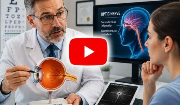
The optic nerve is a bundle of more than one million nerve fibers located at the back of each eye.
Its primary job is to transmit signals from the retina to the brain, where these signals are processed into the images you recognize as vision.
Light enters the eye and reaches the retina, a layer of specialized cells responsible for detecting visual information.
The retina converts light into electrical signals. These signals travel through the optic nerve and reach the visual centers of the brain.
Because the optic nerve connects the eyes and brain, damage to this structure can affect how a person sees.
Problems may influence peripheral vision, color perception, sharpness of vision, or the ability to see clearly in certain areas.
Unlike some tissues in the body, damaged optic nerve fibers have limited ability to repair themselves. This makes early detection and regular monitoring especially important.
Why Do Doctors Test the Optic Nerve?
Eye doctors test the optic nerve to evaluate whether it is functioning normally and whether there are signs of damage or disease.
Many optic nerve problems develop slowly. A person may not notice changes until a significant amount of vision has already been affected.
Regular testing allows doctors to identify warning signs earlier.
Common reasons for optic nerve testing include:
- Monitoring glaucoma risk
- Evaluating unexplained vision changes
- Checking for optic nerve inflammation
- Investigating sudden vision loss
- Examining optic nerve appearance
- Tracking changes over time
Optic nerve examinations are especially important for people with risk factors such as older age, family history of glaucoma, diabetes, high eye pressure, previous eye injuries, or certain neurological conditions.
Signs That May Require Optic Nerve Testing
Some optic nerve conditions may not cause symptoms immediately. However, certain warning signs should be evaluated by an eye care professional.
Possible signs include:
- Blurred or reduced vision
- Loss of side vision
- Difficulty seeing in low light
- Changes in color vision
- Eye pain with vision changes
- Sudden vision loss
- Seeing dark areas or missing spots in vision
- Differences between the vision of both eyes
Sudden vision changes should receive prompt medical attention because some optic nerve conditions can progress quickly.

How to Test the Optic Nerve During an Eye Examination
Eye doctors use multiple tests because the optic nerve has both structural and functional roles.
Some tests examine the physical appearance of the nerve, while others measure how well it sends visual information.
1. Visual Acuity Test
A visual acuity test is one of the most common parts of an eye examination. It measures how clearly a person can see at different distances.
During this test, a patient reads letters, numbers, or symbols from an eye chart while covering one eye at a time.
Although this test does not directly show optic nerve damage, it helps doctors understand overall visual performance and identify changes that may require additional evaluation.
A reduction in visual clarity may lead an eye specialist to perform more detailed optic nerve testing.
2. Pupil Response Test
The pupil response test evaluates how the optic nerve communicates with the brain.
During this examination, a doctor shines a small light into each eye and observes how the pupils react.
A healthy optic nerve helps control normal pupil reactions. If one optic nerve is affected, the doctor may notice differences between the eyes.
This test is commonly used when doctors suspect optic nerve inflammation, nerve injury, or other neurological concerns.
3. Visual Field Test
A visual field test measures a person’s ability to see objects outside the direct line of vision.
The test evaluates peripheral vision, which is often affected by certain optic nerve conditions.
During the procedure, the patient looks straight ahead while responding whenever they see flashes of light in different areas.
Visual field testing is especially important for glaucoma monitoring because glaucoma can gradually damage peripheral vision.
Doctors compare results over time to determine whether vision changes are stable or progressing.
4. Fundus Examination
A fundus examination allows an eye doctor to examine the back of the eye, including the optic nerve head.
The doctor uses special instruments to look through the pupil and evaluate the appearance of structures inside the eye.
During this examination, doctors assess:
- Optic nerve color
- Optic nerve shape
- Nerve fiber appearance
- Signs of swelling
- Changes associated with disease
The appearance of the optic nerve can provide important information about eye health.
5. Optical Coherence Tomography (OCT)
Optical coherence tomography, commonly called OCT, is an advanced imaging test used to create detailed images of the retina and optic nerve area.
OCT uses light waves to capture cross-sectional images of eye structures. It allows doctors to measure the thickness of retinal nerve fiber layers surrounding the optic nerve.
This information helps detect subtle changes that may not be visible during a regular eye examination.
OCT is frequently used for:
- Glaucoma monitoring
- Optic nerve damage assessment
- Tracking changes over time
- Evaluating retinal nerve health
Because OCT provides measurable information, doctors can compare results from different visits.
6. Color Vision Testing
The optic nerve plays an important role in transmitting color information from the eye to the brain.
Color vision testing helps doctors evaluate whether the optic nerve is functioning properly.
During this test, patients identify patterns or numbers within specially designed color plates.
Changes in color perception may occur with certain optic nerve conditions, including inflammation or injury.
7. Eye Pressure Measurement
Eye pressure testing, also known as tonometry, does not directly measure optic nerve function. However, it is an important part of glaucoma evaluation.
High pressure inside the eye can increase stress on the optic nerve and contribute to damage over time.
Doctors combine eye pressure measurements with optic nerve appearance, visual field results, and imaging tests to create a complete assessment.
How Doctors Identify Optic Nerve Damage
Optic nerve damage can appear differently depending on the underlying cause.
Eye specialists evaluate several factors, including:
- Changes in optic nerve structure
- Loss of nerve fiber thickness
- Visual field changes
- Reduced color perception
- Abnormal pupil reactions
A doctor does not rely on one test alone. Combining different examinations provides a clearer understanding of optic nerve health.
For example, a person may have normal vision but show early structural changes on OCT. Another person may have visual field changes that require further investigation.
Optic Nerve Testing for Glaucoma
Glaucoma is one of the most common conditions associated with optic nerve damage.
It occurs when the optic nerve becomes damaged, often related to increased pressure inside the eye. However, glaucoma can also occur with normal eye pressure.
Optic nerve testing helps doctors:
- Detect early changes
- Estimate disease progression
- Monitor treatment effectiveness
- Protect remaining vision
Regular testing is important because glaucoma-related vision loss is often permanent.
Optic Nerve Testing for Optic Neuritis
Optic neuritis is inflammation of the optic nerve that can affect vision.
People with optic neuritis may experience:
- Reduced vision in one eye
- Eye discomfort during movement
- Changes in color brightness
- Blurred vision
Doctors may use vision tests, pupil examinations, imaging, and medical history evaluation to understand the cause.
Some cases require additional neurological evaluation depending on symptoms and findings.
What Happens During an Optic Nerve Examination?
A typical optic nerve evaluation begins with questions about vision changes, medical history, medications, and previous eye problems.
The eye doctor may perform:
- Vision testing
- Pupil examination
- Eye pressure measurement
- Retinal and optic nerve evaluation
- Visual field testing
- Imaging tests if needed
Most tests are painless and usually take a short amount of time.
If abnormal findings appear, the doctor may recommend additional monitoring or treatment depending on the cause.
How Often Should You Test Your Optic Nerve?
The recommended frequency of optic nerve testing depends on age, health history, and individual risk factors.
People with higher risk factors may need more frequent examinations.
Regular eye examinations are important because some optic nerve conditions develop without noticeable symptoms.
Routine testing can help identify changes early and support better long-term vision management.
Factors That Can Affect Optic Nerve Health
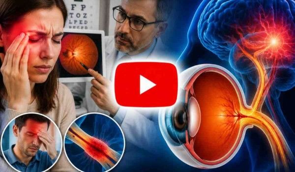
Several factors may influence optic nerve health, including:
➢ Age
The risk of certain optic nerve conditions increases with age. Regular eye examinations become increasingly important as people get older.
➢ Family History
Some eye conditions involving the optic nerve can have genetic factors. A family history of glaucoma may increase the need for monitoring.
➢ Diabetes
Diabetes can affect blood vessels and may contribute to changes affecting vision and eye health.
➢ Eye Injuries
Trauma to the eye area may affect optic nerve function and should be evaluated by an eye professional.
➢ Neurological Conditions
Because the optic nerve connects with the brain, some neurological conditions may influence optic nerve function.
How to Support Long-Term Optic Nerve Health
Protecting overall eye health can help maintain healthy vision.
Helpful habits include:
- Scheduling regular eye examinations
- Managing chronic health conditions
- Protecting eyes from injuries
- Wearing appropriate eye protection when needed
- Following medical recommendations
- Reporting unusual vision changes quickly
Healthy lifestyle choices also support overall circulation and wellness, which are important for eye function.
When Should You See an Eye Doctor?
You should consider professional evaluation if you experience:
- Sudden vision changes
- New blind spots
- Persistent blurred vision
- Eye pain with vision problems
- Reduced peripheral vision
- Unexplained changes in color vision
Early assessment can help determine whether symptoms are related to the optic nerve or another part of the visual system.
Final Thoughts
Understanding how to test the optic nerve helps people recognize the importance of regular eye care and early detection.
The optic nerve is essential for transferring visual information from the eyes to the brain, and protecting its health is a key part of maintaining vision.
Optic nerve testing involves several methods, including visual field exams, pupil response tests, eye imaging, and detailed eye examinations.
Each test provides different information about optic nerve structure and function.
Many optic nerve conditions can be managed more effectively when detected early.
Regular eye checkups, awareness of warning signs, and professional evaluation of vision changes are important steps for maintaining long-term eye health.
References
https://en.wikipedia.org/wiki/Optic_nerve
https://www.healthline.com/health/optic-neuritis
https://www.webmd.com/eye-health
About Us